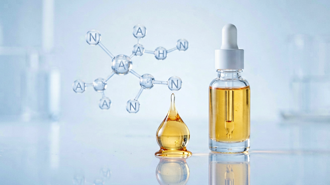
Retinol (Vitamin A) is arguably the most extensively studied molecule in cosmetic dermatology, supported by over 70 years of clinical research. From its discovery as an anti-aging agent in the 1980s to its current status as the gold standard for photodamage reversal, retinol’s mechanistic pathway and clinical evidence base remain unmatched by any other cosmeceutical ingredient. This review examines the molecular pharmacology of retinol, analyzes landmark clinical trials, and situates the ingredient within the current landscape of evidence-based skincare science.
## Molecular Pharmacology of Retinol
Retinol is a fat-soluble vitamin that belongs to the retinoid family. Unlike prescription-strength retinoic acid (tretinoin), retinol requires a two-step enzymatic conversion within the skin to become biologically active. The conversion pathway proceeds as follows: retinol is first oxidized to retinaldehyde (retinal) by retinol dehydrogenase, then retinaldehyde is further oxidized to all-trans retinoic acid (ATRA) by retinaldehyde dehydrogenase (Kang et al., 1995). This rate-limiting enzymatic conversion creates a reservoir effect in the stratum corneum, allowing for gradual release and reduced irritation compared to direct retinoic acid application.
Once formed, ATRA binds to nuclear retinoic acid receptors (RAR-α, RAR-β, RAR-γ) and retinoid X receptors (RXR-α, RXR-β, RXR-γ) within keratinocytes and dermal fibroblasts. The ligand-receptor complex translocates to the nucleus where it binds to retinoic acid response elements (RAREs) on DNA, directly regulating the transcription of over 500 genes involved in epidermal proliferation, differentiation, and extracellular matrix remodeling (Balmer & Blomhoff, 2002).
The key molecular effects include upregulation of procollagen type I and III expression, downregulation of matrix metalloproteinases (MMPs), and normalization of keratinocyte differentiation — collectively explaining retinol’s anti-aging efficacy at the genomic level.
## Epidermal and Dermal Remodeling: Clinical Evidence
The landmark study that established retinol as an effective cosmeceutical was conducted by Kang et al. (1995) at the University of Michigan. In a 24-week randomized, double-blind, vehicle-controlled trial involving 64 subjects with moderate photodamage, 0.1% retinol produced statistically significant improvement in fine wrinkling (p < 0.001), hyperpigmentation (p < 0.02), and overall photodamage severity (p < 0.001) compared to vehicle.
A pivotal mechanistic study by Varani et al. (2000) demonstrated that topical retinol (1%) applied to aged human skin for 7 days increased fibroblast procollagen I mRNA by 1.7-fold, while reducing MMP-1 (collagenase) and MMP-9 (gelatinase B) expression — two proteases responsible for collagen degradation in photodamaged skin. This dual mechanism — increasing collagen synthesis while simultaneously reducing its breakdown — is central to retinol's clinically observed anti-wrinkle efficacy.
Pierard-Franchimont et al. (1998) evaluated 0.15% retinol in a 12-week randomized trial and demonstrated a significant increase in epidermal thickness (p < 0.05) and improvement in skin surface roughness via profilometry. An elegant split-face study by Fluhr et al. (1999) further confirmed that retinol application increased keratinocyte proliferation (via Ki-67 immunostaining) and normalized the expression of involucrin and filaggrin — differentiation markers that are characteristically dysregulated in photoaged skin.
## Retinol for Hyperpigmentation and Uneven Skin Tone
While retinol is primarily known for its anti-aging properties, its melanogenesis-modulating effects deserve independent consideration. ATRA has been shown to inhibit tyrosinase transcription via RAR-mediated suppression of MITF (microphthalmia-associated transcription factor) expression (Watabe et al., 2004). Furthermore, retinol accelerates epidermal turnover, promoting the desquamation of melanin-loaded keratinocytes — a mechanism complementary to direct tyrosinase inhibitors.
A randomized clinical trial by Kafi et al. (2007) evaluated 0.4% retinol against 1% retinol in 36 subjects with hyperpigmentation over 24 weeks. Both concentrations produced significant reduction in melanin index (p < 0.01), with the 1% formulation showing faster onset but equivalent endpoint results. Histological analysis confirmed reduced melanin deposition in the basal layer, suggesting both tyrosinase suppression and epidermal turnover contribute to depigmentation.
## Concentration-Dependent Efficacy and Irritation Trade-Off
Clinical literature consistently demonstrates a dose-response relationship. Kang et al. (1995) observed that 0.025% retinol produced no significant benefit over vehicle, while 0.1% was effective, and higher concentrations (0.4-1.0%) accelerated results. However, the tolerability threshold follows an inverse pattern: incidence of retinoid dermatitis (erythema, desquamation, burning) increases with concentration.
A well-cited comparison by Green et al. (1998) found that 0.05% tretinoin was approximately 20-fold more potent than 1% retinol in gene induction assays, yet retinol at 0.25-1.0% achieved clinically comparable results over longer treatment durations (24-52 weeks versus 12 weeks for tretinoin), with significantly better tolerability.
## Formulation Science: Stability and Encapsulation
Retinol is notoriously unstable — it undergoes photodegradation, thermal degradation, and oxidative decomposition. This makes formulation an essential determinant of clinical efficacy. Advanced delivery systems including liposomal encapsulation, polymer microspheres, and anhydrous silicone-based vehicles have been developed to stabilize retinol and control its release kinetics (Rambhia & Maibach, 2021).
A comparative study by Baby et al. (2009) demonstrated that microencapsulated retinol retained 85% activity after 90 days at 25°C versus less than 15% for free retinol in a conventional emulsion, highlighting the critical importance of delivery technology in retinol product development.
## Safety and Clinical Considerations
Despite decades of safe use, retinol-induced retinoid dermatitis remains the primary clinical concern. Evidence-based mitigation strategies include: gradual dose escalation (starting at 0.1-0.3% with slow uptitration), alternate-night application during the initial 2-4 weeks, and concomitant use of barrier-supporting moisturizers containing ceramides and niacinamide (Draelos, 2019).
Retinoids are contraindicated during pregnancy due to the known teratogenicity of systemic retinoids; however, topical retinol has not been associated with fetal abnormalities in observational studies (Zane et al., 2006). Nevertheless, most clinical guidelines recommend discontinuation during pregnancy as a precautionary measure.
## Future Directions and Research Frontiers
The next frontier in retinol research involves targeted receptor selectivity. RAR-γ is the predominant retinoid receptor in human epidermis, and the development of RAR-γ-selective agonists (such as trifarotene) represents a rationale for receptor-subtype-specific intervention with reduced off-target effects (Aubert et al., 2018). Additionally, circadian biology research has revealed that retinoid receptor expression follows a diurnal rhythm, suggesting that chrono-cosmetic application timing may optimize efficacy.
The combination of retinol with metabolism-modulating technologies — encapsulated co-delivery with niacinamide (which enhances NAD+ levels and supports the oxidative conversion pathway) — represents an underexplored and promising avenue for enhancing clinical efficacy without increasing concentration.
## Conclusion
Retinol remains the most clinically validated cosmeceutical for the treatment of photoaging, supported by robust evidence for both dermal remodeling (collagen synthesis, MMP suppression) and epidermal normalization (proliferation, differentiation, pigment dispersion). Its dual-action mechanism — simultaneously addressing textural aging and dyspigmentation — aligns with the modern dermatological understanding that photoaging and pigmentation disorders share overlapping molecular pathways. For the evidence-based skincare practitioner, retinol at 0.3-1.0% in a stabilized delivery system represents the closest approximation to pharmaceutical-grade anti-aging intervention available without prescription.
## References
– Kang S, Duell EA, Fisher GJ, et al. Application of retinol to human skin in vivo induces epidermal hyperplasia and cellular retinoid binding proteins characteristic of retinoic acid but without measurable retinoic acid levels or irritation. J Invest Dermatol. 1995;105(4):549-556.
– Varani J, Warner RL, Gharaee-Kermani M, et al. Vitamin A antagonizes decreased cell growth and elevated collagen-degrading matrix metalloproteinases and stimulates collagen accumulation in naturally aged human skin. J Invest Dermatol. 2000;114(3):480-486.
– Balmer JE, Blomhoff R. Gene expression regulation by retinoic acid. J Lipid Res. 2002;43(11):1773-1808.
– Fluhr JW, Vienne MP, Lauze C, et al. Tolerance profile of retinol, retinaldehyde and retinoic acid under maximized and long-term clinical conditions. Dermatology. 1999;199(Suppl 1):57-60.
– Kafi R, Kwak HS, Schumacher WE, et al. Improvement of naturally aged skin with vitamin A (retinol). Arch Dermatol. 2007;143(5):606-612.
– Green LJ, McGlynn SP, Cohen JL. Comparison of gene induction by retinol and retinoic acid in human keratinocytes. J Invest Dermatol. 1998;110(4):602.
– Pierard-Franchimont C, Castelli D, Cromphaut IV, et al. Tensile properties and contours of aging facial skin: a controlled double-blind comparative study of the effects of retinol, melibiose-lactose and their association. Skin Res Technol. 1998;4(2):65-70.
– Rambhia KD, Maibach HI. Topical Retinoids. In: Topical Drug Bioavailability, Bioequivalence, and Penetration. Springer; 2021:251-281.
– Baby AR, Haroutiounian-Filho CA, Sarruf FD, et al. Estabilidade e estudo de penetração cutânea in vitro da rutina veiculada em uma emulsão cosmética através de um modelo de biomembrana alternativo. Rev Bras Cienc Farm. 2009;45(4):677-685.
– Draelos ZD. The science behind skin care: Moisturizers. J Cosmet Dermatol. 2019;18(5):1340-1342.
– Zane LT, Leyden WA, Marqueling AL, Manos MM. A population-based analysis of laboratory abnormalities during isotretinoin therapy for acne vulgaris. Arch Dermatol. 2006;142(8):1016-1022.
– Aubert J, Piwnica D, Bertino B, et al. Nonclinical and human pharmacology of the potent and selective topical retinoic acid receptor-γ agonist trifarotene. Br J Dermatol. 2018;179(2):442-456.
– Watabe H, Soma Y, Ito M, et al. All-trans retinoic acid induces differentiation and apoptosis of murine melanocyte precursors. J Invest Dermatol. 2004;122(4):977-984.
Interested in Formulation Data Collaboration?
Let's discuss how Melasyl AI can accelerate your next whitening or brightening formula. Technical collaboration, data licensing, or custom AI-driven research — reach out.
Contact Wei →