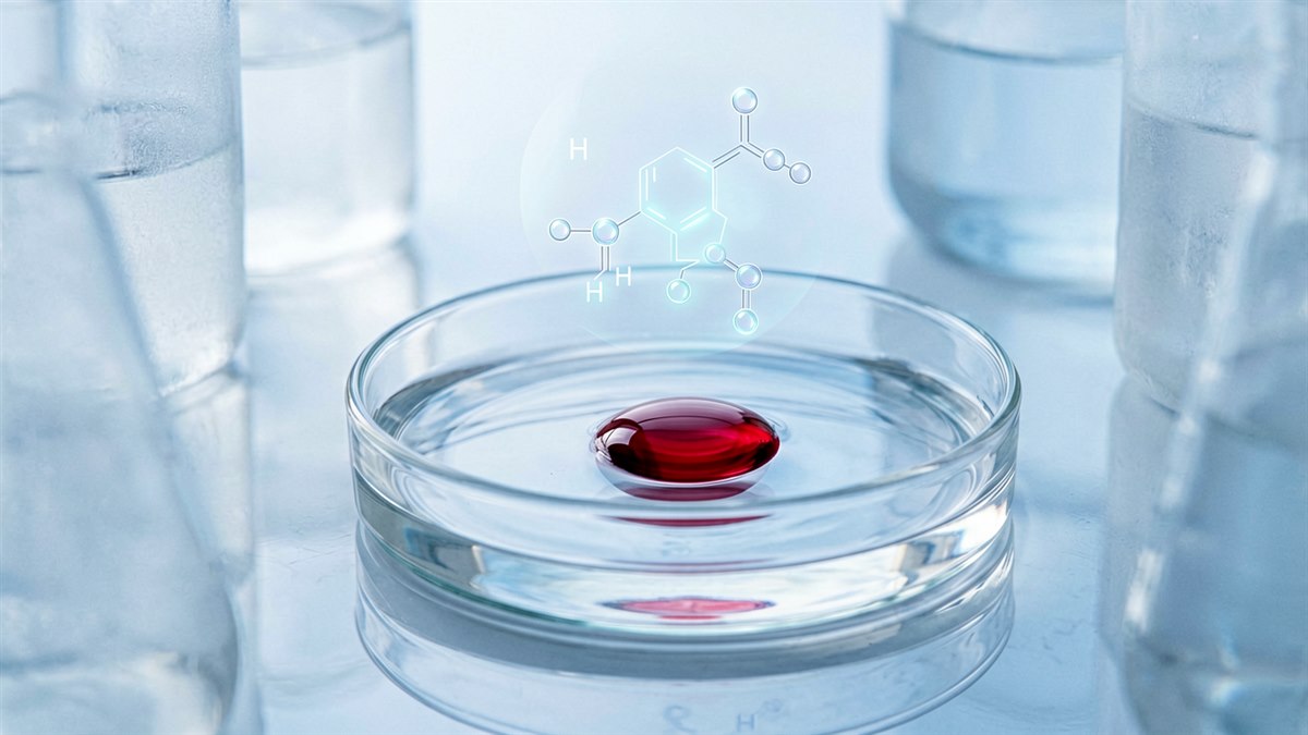
Astaxanthin for hyperpigmentation occupies an unusual position in brightening science: it is one of the few pigment-active ingredients whose effect is not primarily explained by tyrosinase inhibition. While the industry has spent a decade optimising direct enzyme blockers — arbutin, thiamidol, resorcinol derivatives — a parallel body of Japanese cell-biology research has shown that this red xanthophyll carotenoid acts one step upstream, silencing the keratinocyte-to-melanocyte signals that switch melanogenesis on in the first place.
That distinction explains why astaxanthin’s clinical file looks contradictory when read carelessly. This review separates the mechanistic evidence from the human outcome data.
What Astaxanthin Actually Is
Astaxanthin (INCI: Astaxanthin; CAS 472-61-7) is a keto-carotenoid, C40H52O4, MW 596.84 g/mol, sourced commercially from the microalga Haematococcus pluvialis. Structurally it is a xanthophyll: a conjugated polyene backbone terminated by two ionone rings, each carrying both a hydroxyl and a ketone group.
That terminal oxygenation is the entire story. Unlike beta-carotene, astaxanthin’s polar end groups let the molecule span the lipid bilayer, anchoring at both membrane surfaces while the conjugated chain sits in the hydrophobic core. It therefore quenches singlet oxygen precisely at the membrane interface where lipid peroxidation initiates, rather than partitioning uselessly into the bilayer centre.
Rao et al. (2013) compared free astaxanthin against the mono- and di-esters native to H. pluvialis in a UV/DMBA rodent model: the esters normalised an approximately 7-fold tyrosinase elevation where the free form managed only 1.4-2.2-fold, most plausibly through better bioavailability.
Mechanism: Interrupting the Paracrine Pigmentation Loop
UV-induced pigmentation is not a melanocyte-autonomous event. Irradiated keratinocytes secrete melanogenic cytokines — endothelin-1 (ET-1), stem cell factor (SCF), GM-CSF, IL-1-alpha, IL-6 and IL-8 — which drive MITF expression and tyrosinase transcription in neighbouring melanocytes. Any active that blunts that secretion reduces pigment without touching the enzyme.
Niwano et al. (2015), in Cytokine, built a keratinocyte-melanocyte insert co-culture to interrogate this loop. UVB exposure of the keratinocyte compartment raised melanocyte MITF, tyrosinase and TRP-1 gene expression and increased tyrosinase catalytic activity. An ET-1 neutralising antibody abolished the tyrosinase rise, confirming ET-1 as the dominant driver.
Two findings are formulation-relevant. First, astaxanthin applied after irradiation still abolished the UVB-induced tyrosinase up-regulation, and significantly reduced secreted ET-1, GM-CSF, IL-1-alpha and IL-6/8. Second, astaxanthin did not block tyrosinase activity when ET-1 was applied directly to melanocytes — a clean demonstration that its action sits at the keratinocyte signal source, not the melanocyte receptor cascade.
The MSK1 Node
A second target does sit inside the melanocyte. Niwano et al. (2018), in Archives of Dermatological Research, showed that SCF stimulates melanogenesis through a previously uncharacterised c-KIT-dependent p38/MSK1 branch feeding into CREB phosphorylation and MITF induction. Astaxanthin left p38 activation intact but suppressed phosphorylation of its downstream target MSK1 — and with it CREB activation and MITF protein at 1.5 hours, followed by reduced tyrosinase and endothelin B receptor protein at 96 hours. Silencing MSK1 reproduced the effect, confirming the node.
Imokawa’s 2019 synthesis in Photochemistry and Photobiology frames the consequence bluntly: these effects are reactive-oxygen-species-depletion independent. Astaxanthin is acting as a kinase-pathway modulator, not a radical sponge. Post-irradiation efficacy is the practical signature of that mechanism — most antioxidants must be present before the insult.
Direct enzyme inhibition is the weakest part of the file. In vitro anti-tyrosinase activity is reported, but potency is modest and unlikely to contribute at realistic topical use levels — marketing astaxanthin as a tyrosinase inhibitor sets up a claim that will not survive scrutiny.
What the Human Evidence Supports — and What It Does Not
Tominaga et al. (2012), Acta Biochimica Polonica. The most-cited pigmentation-relevant study: 30 healthy women, 8 weeks, 6 mg/day oral plus 2 mL/day topical astaxanthin (78.9 micromolar). Reported reduced age spot size on the cheek at week 8, alongside wrinkle, elasticity and corneocyte gains. It is open-label and uncontrolled — hypothesis-generating, not confirmatory. The companion arm (36 men, randomised, double-blind, placebo-controlled, 6 weeks) improved crow’s feet and transepidermal water loss but reported no pigment endpoint.
Ito et al. (2018), Nutrients. Methodologically the strongest single trial: 23 Japanese adults, 10 weeks, randomised double-blind placebo-controlled, 4 mg/day. The astaxanthin arm showed an increased minimal erythema dose (MED) and reduced UV-induced moisture loss in irradiated skin. A raised MED is mechanistically relevant to pigmentation prevention, since erythemal threshold and melanogenic stimulation share upstream inflammatory mediators — but MED is a photoprotection endpoint, not a melanin index.
Zhou et al. (2021), Nutrients. Meta-analysis of 11 studies: oral astaxanthin improved moisture (SMD 0.53; 95% CI 0.05-1.01) and elasticity (SMD 0.77; 95% CI 0.19-1.35), but not wrinkle depth, with high heterogeneity for elasticity (I-squared 75%). Ng et al. (2021) flagged the recurring limitations: small samples, predominantly healthy Japanese women, and frequent commercial sponsorship.
Yang et al. (2025), Frontiers in Medicine. The most recent word is deflationary. Pooling 40 randomised controlled trials of oral supplements for photoaging, the authors concluded there is insufficient evidence to recommend astaxanthin — in contrast to collagen and flavanols, which did shift MED or elasticity.
For topical delivery, Konisky et al. (2023) reported improved radiance and even tone over 12 weeks in 32 women — but the test article combined vitamin C, astaxanthin, fermented turmeric and vitamin E, and the study was open-label, so no share of that effect can be attributed to astaxanthin.
Evidence Grading Summary
| Claim | Best evidence | Grade |
|---|---|---|
| Blocks UVB-induced paracrine melanogenic signalling | Co-culture, post-irradiation, ET-1 quantified | Strong (in vitro) |
| Suppresses MSK1/CREB/MITF axis | Human melanocytes, MSK1 silencing control | Strong (in vitro) |
| Raises MED, reduces UV barrier loss | Randomised, double-blind, placebo-controlled | Moderate (small n) |
| Improves moisture and elasticity (oral) | Meta-analysis, 11 studies | Moderate |
| Reduces age spot size in humans | Open-label, uncontrolled, combined route | Weak |
| Direct tyrosinase inhibition at use levels | In vitro only, modest potency | Weak |
Formulation Reality
Colour load. At functional levels — roughly 0.01-0.05% pure astaxanthin — the product is unavoidably orange-red, and above about 0.05% transient staining on light textiles and fair skin becomes a genuine complaint driver. A deliberate amber-to-coral shade story is easier than fighting the chromophore.
Oxidative stability. The conjugated chain that quenches singlet oxygen is itself readily oxidised, so encapsulation is effectively mandatory: oleoresin in tocopherol-stabilised MCT, liposomal systems, or solid lipid nanoparticles. Package opaque and airless, and run 40 degrees C accelerated testing with colorimetric measurement alongside pH — colour fade precedes measurable potency loss.
Positioning. Because the documented action is prevention of paracrine signalling rather than correction of an existing melanin load, astaxanthin belongs in daytime antioxidant and post-sun repair products layered with sunscreen — not as the hero active in a dark-spot treatment. If correction is the claim, pair it with a validated tyrosinase inhibitor.
Summary
Astaxanthin for hyperpigmentation has one of the most mechanistically interesting and clinically underpowered evidence bases in brightening science. The cell biology is unusually well characterised: it interrupts UVB-driven keratinocyte cytokine secretion, works after irradiation, and blocks the p38/MSK1/CREB/MITF axis independently of radical scavenging. That is a genuinely differentiated mechanism in a category saturated with enzyme inhibitors.
The human pigment data has not caught up. The only study reporting age spot reduction was open-label and combined oral with topical dosing, and the 2025 pooled analysis declined to recommend oral astaxanthin. The honest position: a well-supported photoprotective antioxidant with a strong mechanistic rationale for pigmentation prevention, and an unproven record for correction. The gap is a properly powered randomised topical trial using melanin index as the primary endpoint in Fitzpatrick III-V skin.
References
- Niwano T, Terazawa S, Nakajima H, Wakabayashi Y, Imokawa G. Astaxanthin and withaferin A block paracrine cytokine interactions between UVB-exposed human keratinocytes and human melanocytes via the attenuation of endothelin-1 secretion and its downstream intracellular signaling. Cytokine. 2015;73(2):184-197. PMID: 25777483.
- Niwano T, Terazawa S, Nakajima H, Imokawa G. The stem cell factor-stimulated melanogenesis in human melanocytes can be abrogated by interrupting the phosphorylation of MSK1: evidence for involvement of the p38/MSK1/CREB/MITF axis. Arch Dermatol Res. 2018;310(3):187-196. PMID: 29362867.
- Imokawa G. Intracellular signaling mechanisms involved in the biological effects of the xanthophyll carotenoid astaxanthin to prevent the photo-aging of the skin in a reactive oxygen species depletion-independent manner. Photochem Photobiol. 2019;95(2):480-489. PMID: 30317634.
- Imokawa G. The xanthophyll carotenoid astaxanthin has distinct biological effects to prevent the photoaging of the skin even by its postirradiation treatment. Photochem Photobiol. 2019;95(2):490-500. PMID: 30338860.
- Ito N, Seki S, Ueda F. The protective role of astaxanthin for UV-induced skin deterioration in healthy people — a randomized, double-blind, placebo-controlled trial. Nutrients. 2018;10(7):817. PMID: 29941810.
- Tominaga K, Hongo N, Karato M, Yamashita E. Cosmetic benefits of astaxanthin on human subjects. Acta Biochim Pol. 2012;59(1):43-47. PMID: 22428137.
- Zhou X, Cao Q, Orfila C, Zhao J, Zhang L. Systematic review and meta-analysis on the effects of astaxanthin on human skin ageing. Nutrients. 2021;13(9):2917. PMID: 34578794.
- Ng QX, De Deyn MLZQ, Loke W, Foo NX, Chan HW, Yeo WS. Effects of astaxanthin supplementation on skin health: a systematic review of clinical studies. J Diet Suppl. 2021;18(2):169-182. PMID: 32202443.
- Yang Q, Li H, Zhang H, Ma L, Zhang X, Wu J. Effectiveness of dietary supplements for skin photoaging in healthy adults: a systematic review and meta-analysis of randomized controlled trials. Front Med (Lausanne). 2025;12:1582946. PMID: 40761858.
- Rao AR, Sindhuja HN, Dharmesh SM, Sankar KU, Sarada R, Ravishankar GA. Effective inhibition of skin cancer, tyrosinase, and antioxidative properties by astaxanthin and astaxanthin esters from the green alga Haematococcus pluvialis. J Agric Food Chem. 2013;61(16):3842-3851. PMID: 23473626.
- Konisky H, Bowe WP, Yang P, Kobets K. A clinical evaluation of the efficacy and tolerability of a novel topical antioxidant formulation featuring vitamin C, astaxanthin, and fermented turmeric. J Cosmet Dermatol. 2023;22(11):3088-3094. PMID: 37608511.
Interested in Formulation Data Collaboration?
Let's discuss how Melasyl AI can accelerate your next whitening or brightening formula. Technical collaboration, data licensing, or custom AI-driven research — reach out.
Contact Wei →