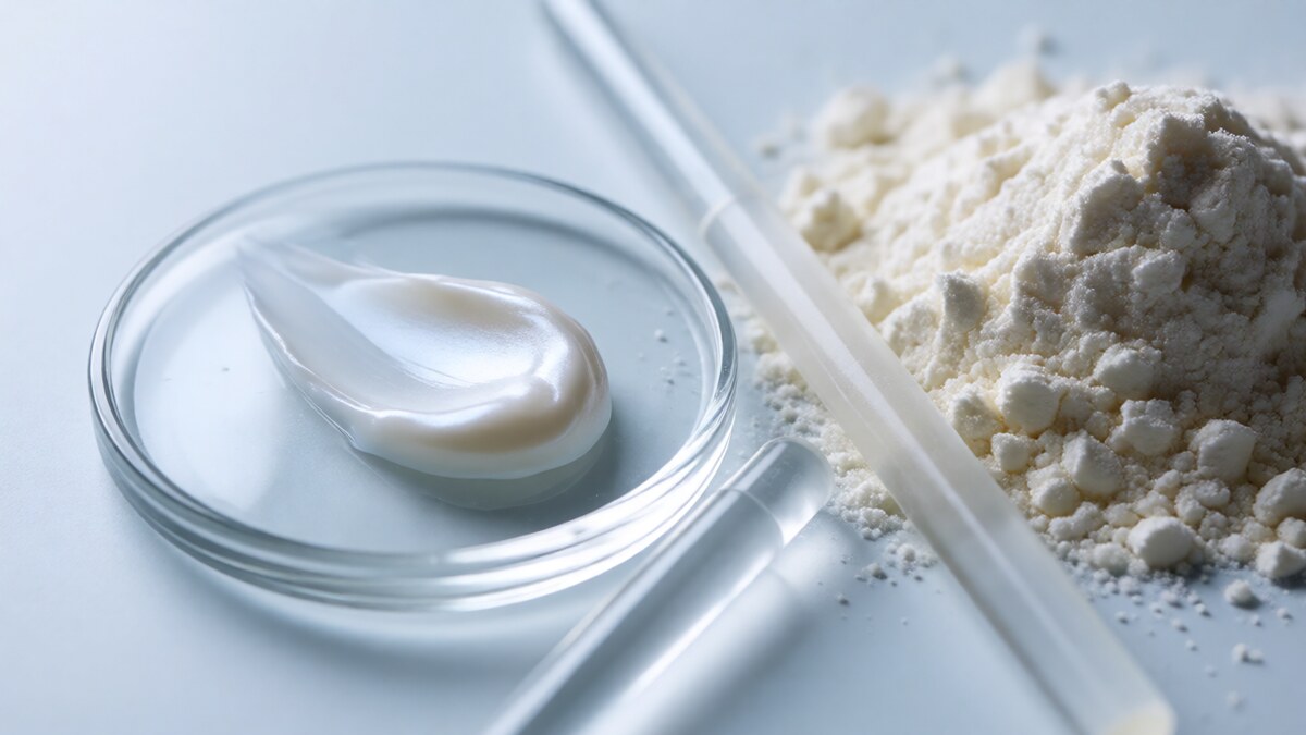
Why an Iron-Binding Milk Protein Became a Hyperpigmentation Story
Most brightening actives attack the same node: the tyrosinase enzyme. Lactoferrin is different. It is a transferrin-family glycoprotein found in milk, tears, saliva and neutrophil granules, and its native job is to sequester free iron. That single property — iron chelation — is why it has quietly entered the dyschromia conversation, and it is also why the mechanistic literature around it reads unlike anything in the standard tyrosinase-inhibitor stack.
The skin-relevant argument runs on two rails. The first is iron. Free ferrous iron drives the Fenton reaction, generating hydroxyl radicals that both amplify oxidative stress and feed the inflammatory signalling that pushes melanocytes into overdrive. By tightly binding labile iron, lactoferrin removes the fuel. The second rail is direct transcriptional control of the pigmentation programme. This article reviews what the studies actually show, where the evidence is strong, and where the marketing has run ahead of the data.
Mechanism One: Iron Chelation and the Pigment Precursor Problem
Iron is not a bystander in pigmentation. Tyrosinase is a copper-dependent enzyme, but the oxidative environment that supports melanogenesis depends on redox-active iron. Iron released from broken capillaries is also the substrate for hemosiderin, the brown pigment behind bruising and a subset of stubborn facial discoloration. Lactoferrin’s iron-binding sites directly limit both pathways: less Fenton chemistry, less hemosiderin deposition, and a calmer inflammatory milieu.
This is the mechanism behind lactoferrin’s most consistent clinical use — post-procedure bruising and redness. A topical formulation combining lactoferrin with arnica and other anti-inflammatory agents has been shown in controlled work to accelerate recovery and reduce bruising after dermal filler injections. For pigmentation specialists, that same iron-sequestration logic is the bridge to treating inflammatory and vascular-pattern dyschromia, not just classic epidermal melasma.
Mechanism Two: The ERK-MITF Shutdown
The more surprising finding is that lactoferrin actively suppresses melanogenesis rather than simply removing oxidative triggers. In a widely cited 2017 study published in Biochemistry and Cell Biology, Ishii, Ryu and Suzuki (Saraya Co. Lactoferrin Laboratory) tested bovine lactoferrin on a three-dimensional cultured pigmentation skin model and in human melanoma cells. Two results matter:
- In the 3D pigmented skin model, lactoferrin produced roughly a 20% reduction in pigmentation, evidence that it was transdermally absorbed and suppressed melanin production.
- In melanocytes, lactoferrin caused a significant, dose-dependent suppression of melanin synthesis.
The mechanism was traced to microphthalmia-associated transcription factor (MITF), the master regulator of the pigmentation gene programme. Lactoferrin both reduced MITF mRNA expression and enhanced degradation of the MITF protein. The primary effector was increased phosphorylation of extracellular signal-regulated kinase (ERK), which tags MITF for degradation and thereby collapses tyrosinase activity downstream. Notably, iron-loaded (holo) and iron-free (apo) lactoferrin suppressed melanogenesis to the same degree — a key control showing the anti-melanogenic action is not purely an iron-starvation artifact but an intrinsic signalling effect.
What the Clinical Evidence Actually Shows
The cell and tissue data are strong and mechanistically coherent. Human clinical data are thinner, and it is important to separate them.
Facial dyschromia in a multi-active formula. The best-controlled human evidence comes from a multi-centre, randomized, blinded trial of a topical product (Alastin PATH-3 technology) that lists lactoferrin among several pigment-pathway actives, alongside hexapeptide-11, phosphatidylserine and tranexamic acid. The pivotal trial (Wang et al., Journal of Drugs in Dermatology, 2023) reported improvements in facial dyschromia, and the 24-week extension phase (Widgerow et al., 2024) showed continued improvement in mMASI and skin evenness with good tolerability. The critical caveat: because the product is a blend, the trial cannot isolate lactoferrin’s individual contribution. It supports the combination strategy, not lactoferrin monotherapy.
Acute and chronic cutaneous dyschromia. An observational study (Musso et al., Acta Vulnologica, 2015) followed 50 patients using a topical lactoferrin preparation. Dyschromia resolved in all acute cases — post-traumatic and post-surgical — typically within the first month, with no intolerance reported even under prolonged use. In chronic dyschromia from underlying venous pathology, results were modest. This pattern is consistent with the iron/hemosiderin mechanism: lactoferrin performs best where the discoloration is inflammatory or vascular in origin.
Sebum and acne support. In vitro work on SZ95 sebocytes showed lactoferrin (10–500 µg/mL) reduced lipid content and downregulated SREBP-1, FAS, SCD-1 and the inflammatory cytokine IL-8. An open-label German study of 43 participants with mild-to-moderate facial acne given 100 mg oral bovine lactoferrin twice daily for eight weeks reported improvement. This matters for pigmentation because acne is a leading driver of post-inflammatory hyperpigmentation in darker skin phototypes.
The Vendor Claim Worth Flagging
Supplier material frequently states that 0.5% lactoferrin matches the melanogenesis inhibition of 2% tranexamic acid in a 3D skin model. That comparison originates from manufacturer research, not an independent peer-reviewed head-to-head trial, and it uses a single model system. Treat it as a directional signal about potency, not as established equivalence. The same caution applies to the “twice as effective as vitamin C for sun-related damage” framing — useful for positioning, not yet backed by controlled clinical comparison.
Formulation and the Translation Gap
Lactoferrin is a large (~80 kDa) glycoprotein, so the honest questions are stability and penetration. The 3D-model data confirm transdermal absorption is achievable, but formulation choices decide whether that holds in a finished product.
- Stability. Lactoferrin is relatively robust to processing but is a protein — avoid prolonged high-temperature phases and strongly denaturing conditions. Its iron-binding pocket and tertiary structure underpin activity.
- pH and chelation. Formulate within a range that preserves iron-binding. Avoid chelator competition from excess EDTA and be deliberate about other metal ions in the system.
- Penetration. Pair with delivery systems that support protein permeation; the effect is dose- and exposure-dependent.
- Preservation. Protein actives need preservative systems chosen for compatibility, not just broad-spectrum kill.
- Positioning. Given the mechanism, lactoferrin is most defensible in post-procedure recovery, bruising, inflammatory PIH and combination dyschromia formulas rather than as a solo melanin-blocker.
The Bottom Line
Lactoferrin is a genuinely differentiated hyperpigmentation active: it removes the redox fuel for pigmentation via iron chelation and simultaneously shuts down the ERK-MITF axis that drives tyrosinase expression. The mechanistic evidence is solid and reproducible, and the clinical signal is strongest in inflammatory, vascular and post-procedure dyschromia — exactly the presentations that resist pure tyrosinase inhibitors. The gap is standalone, adequately powered, ingredient-isolated human trials. Until those exist, the credible claim is a well-supported adjunct with a distinct mechanism, not a hydroquinone replacement.
Key Citations
- Ishii N, Ryu M, Suzuki YA. Lactoferrin inhibits melanogenesis by down-regulating MITF in melanoma cells and normal melanocytes. Biochem Cell Biol. 2017;95(1):119–125. doi:10.1139/bcb-2016-0053.
- Wang J, Fabi S, Robinson D, et al. A multi-center, randomized, blinded clinical study evaluating the efficacy and safety of a novel topical product for facial dyschromia. J Drugs Dermatol. 2023;22(4):333–338.
- Widgerow A, et al. Extension-phase analysis of a topical PATH-3 dyschromia product, 24 weeks. J Drugs Dermatol. 2024;23(1). doi:10.36849/JDD.7622.
- Musso A, Aloesio R, Caponi R. Use of a topical preparation based on lactoferrin to manage acute and chronic cutaneous dyschromia and hypotrophy. Acta Vulnologica. 2015;13(2):77–89.
Interested in Formulation Data Collaboration?
Let's discuss how Melasyl AI can accelerate your next whitening or brightening formula. Technical collaboration, data licensing, or custom AI-driven research — reach out.
Contact Wei →