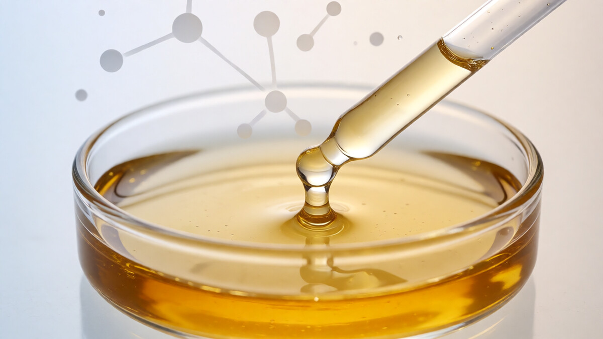
Manuka honey has moved from the wound-care clinic into the brightening aisle. In 2026, honey from the New Zealand mānuka bush (Leptospermum scoparium) is the headline active in dark-spot masks, overnight repair creams and “bee-derived” glow serums — the newest member of a bee-ingredient cluster that already includes royal jelly and bee venom. This analysis breaks down what the literature actually supports for hyperpigmentation, and where the marketing outruns the data.
What Is the Active? A Multi-Compound Fraction
Mānuka honey is not a single molecule, and that is the source of both its appeal and its ambiguity. Three fractions drive the pigment story:
- Methylglyoxal (MGO) — the signature compound that distinguishes mānuka from ordinary honey, present at concentrations 100–1000× higher than in conventional honeys (Mavric et al., Mol Nutr Food Res, 2008). MGO is primarily an antimicrobial, but by suppressing Cutibacterium acnes colonisation it indirectly reduces the inflammatory load that drives post-inflammatory hyperpigmentation (PIH).
- Polyphenols and flavonoids — quercetin, ellagic acid, gallic acid and caffeic acid. These are the direct pigment-relevant actives: several are documented tyrosinase inhibitors that chelate the copper centre of the enzyme.
- Enzymatic hydrogen peroxide and gluconic acid — generated by glucose oxidase as the honey is diluted by skin moisture. Gluconic acid is a mild alpha-hydroxy acid that accelerates surface turnover and lifts dull, melanin-laden corneocytes.
The practical implication is that the pigment benefit is spread across several mechanisms rather than concentrated in one “hero” molecule — which makes standardised, formulated delivery more important than the raw jar.
Mechanism: Three Converging Pathways
1. Anti-inflammatory suppression of melanocyte stimulation
Post-inflammatory hyperpigmentation is driven less by UV than by the cytokine cascade that follows inflammation: IL-1α, endothelin-1 and stem cell factor all stimulate neighbouring melanocytes to overproduce melanin. Honey’s anti-inflammatory activity therefore attacks the upstream cause of PIH. In the best-controlled human study to date, Alangari et al. (Immunity, Inflammation and Disease, 2017) showed that mānuka honey down-regulated IL-4-induced CCL26 release from HaCaT keratinocytes in a dose-dependent manner and significantly inhibited mast cell degranulation — two measurable brakes on the inflammatory signalling that seeds pigmentation.
2. Direct tyrosinase inhibition by polyphenols
Honey’s phenolic fraction — dominated by quercetin and ellagic acid — inhibits tyrosinase through copper chelation and by scavenging the reactive oxygen species that amplify melanogenesis. This is a weaker, dose-dependent effect than kojic acid or arbutin in cell-free assays, but it operates in the same layer as the anti-inflammatory action and complements it. Reviews of natural depigmenting agents place honey polyphenols in the “moderate evidence” tier: solid laboratory activity, limited large human trials.
3. Barrier and acid-mantle support
Mānuka honey sits at pH 3.2–4.5, close to the skin’s natural acid mantle. That mildly acidic environment favours keratinocyte turnover and discourages the barrier disruption that itself triggers melanocyte activation. Honey is also strongly hygroscopic, drawing water into the stratum corneum — useful in the dehydrated, post-acne skin where PIH is most persistent.
Clinical Evidence: What the Trials Show
Atopic dermatitis (mechanistic anchor). Alangari et al. (2017) applied mānuka honey to one of two bilaterally symmetric lesions in 14 adults for seven consecutive nights, using the contralateral lesion as an internal control. Treated lesions improved significantly on the Three Item Severity score versus controls, with no significant adverse events. The trial is small and open-label, but the within-patient design and the corroborating cellular data make it the strongest mechanistic evidence for honey’s inflammation-pigment axis.
Hyperpigmentation and dark spots. O’Gorman et al. (Cureus, 2025) followed 40 women aged 40–55 using a mānuka honey-based serum containing royal jelly and bee venom twice daily for eight weeks. Self-reported dark-spot scores improved significantly from week 2 and were sustained to week 8, and dermatologist grading found improved skin brightness in 60.6% of participants. Because the formula combined three bee actives, the result supports the combination rather than honey alone — an honest limitation the authors acknowledge.
Acne (negative control). A randomised trial of medical-grade kanuka honey — a close mānuka relative — found no significant benefit over comparator for acne lesions. This is the counterweight worth citing: honey’s antibacterial action does not translate into acne clearance on its own, so its pigment value rests on the anti-inflammatory and polyphenol pathways, not on antibacterial claims.
Formulation Guidance for 2026
- Standardise, do not dilute. Retail honey varies enormously in MGO and polyphenol content. Use a standardised mānuka extract (MGO 500+ / UMF 15+ equivalent) so batch-to-batch potency is predictable.
- Preserve the polyphenols. Heat and prolonged processing degrade the flavonoids and glucose oxidase. Formulate with cold-processed extract and avoid high-temperature emulsification.
- Pair for synergy. Honey is a supporting act, not a monotherapy. Combining it with niacinamide and NAG for melanosome transfer control, or a tyrosinase inhibitor such as alpha arbutin, builds a multi-pathway brightening stack.
- Respect the pH window. Keep finished formula pH in the 4.0–5.5 range to preserve honey’s acid-mantle benefit without destabilising co-actives.
- Sunscreen is non-negotiable. Any PIH protocol without daily SPF will be outrun by new pigment formation.
Bottom Line
Mānuka honey is a credible but modest brightening active. Its strongest pigment-relevant mechanism is anti-inflammatory — it calms the cytokine cascade that drives PIH — supported by polyphenol tyrosinase inhibition and gentle acid-mantle turnover. The human data are small and mostly from combination formulas, so it belongs in a supporting role within a multi-pathway routine, not as a stand-alone dark-spot cure. For formulators building 2026 bee-derived brightening lines, it is the complementary anti-inflammatory anchor to royal jelly’s MITF suppression and bee venom’s melittin-driven pathway.
References
- Alangari AA, Morris K, Lwaleed BA, et al. Honey is potentially effective in the treatment of atopic dermatitis: clinical and mechanistic studies. Immunity, Inflammation and Disease. 2017;5(2):190-199. doi:10.1002/iid3.153
- O’Gorman E, Varanasi S, Bukoski S, et al. Investigating the effects of a mānuka honey, royal jelly, and bee venom-derived face serum on skin health and signs of aging. Cureus. 2025;17(3):e81244. doi:10.7759/cureus.81244
- Mavric E, Wittmann S, Barth G, Henle T. Identification and quantification of methylglyoxal as the dominant antibacterial constituent of Leptospermum scoparium (mānuka) honeys. Molecular Nutrition & Food Research. 2008;52(4):483-489.
- Carter DA, Blair SE, Cokcetin NN, et al. Therapeutic mānuka honey: no longer so alternative. Frontiers in Microbiology. 2016;7:569.
- Semprini A, Braithwaite I, Corin A, et al. Randomised controlled trial of topical kanuka honey for the treatment of acne. BMJ Open. 2016;6(2):e009888.
Interested in Formulation Data Collaboration?
Let's discuss how Melasyl AI can accelerate your next whitening or brightening formula. Technical collaboration, data licensing, or custom AI-driven research — reach out.
Contact Wei →