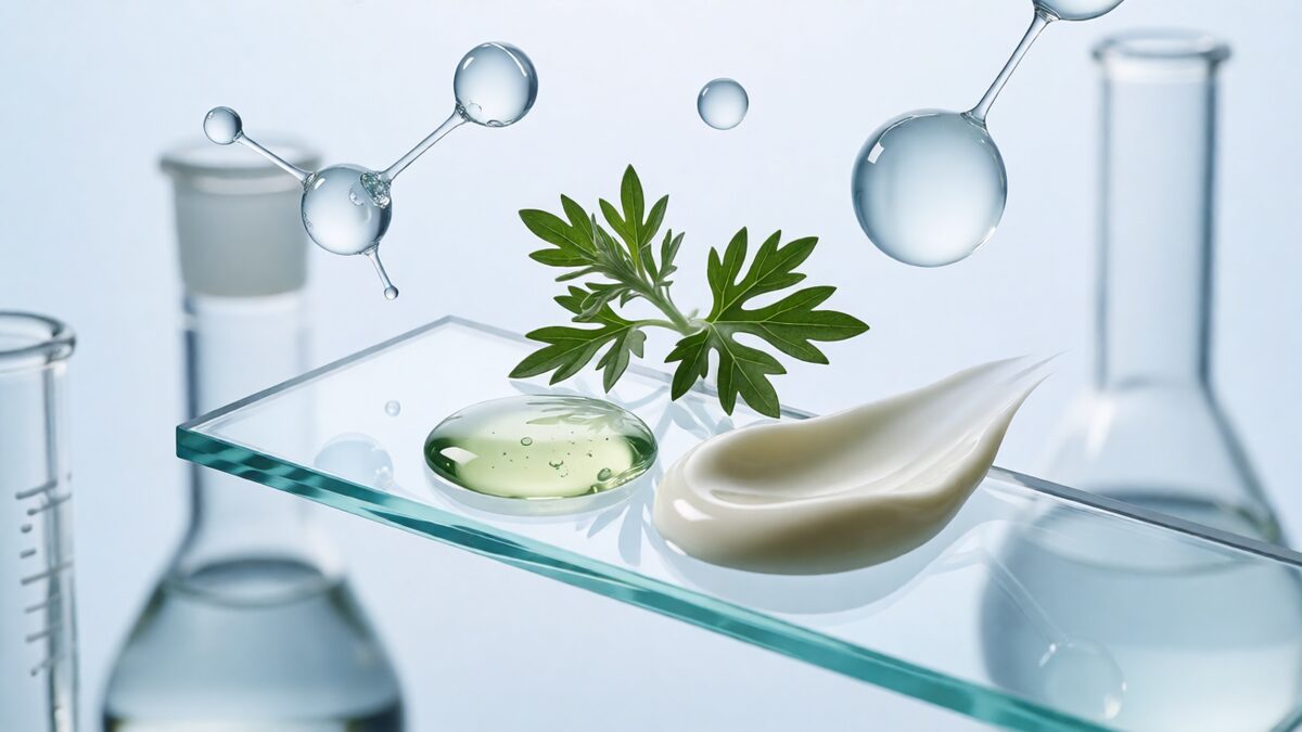
Mugwort has spent years as K-beauty’s signature calming botanical — the hero extract in bestsellers like I’m From Mugwort Essence, Round Lab’s Mugwort Calming Toner and Missha’s Time Revolution Artemisia Treatment Essence. What most ingredient lists never advertise is that the same plant carries a documented, mechanism-level case for hyperpigmentation. The science points to a family of caffeoylquinic acids that suppress melanin not by blocking tyrosinase directly, but by dismantling the transcription factor that commands it.
This analysis separates the marketing from the evidence: which Artemisia species actually brighten, which molecule does the work, what the clinical record supports, and where the formulation risks sit.
Species matters: “mugwort” is not one plant
Mugwort is a common name covering dozens of Artemisia species, and their chemistry diverges enough that they are not interchangeable actives:
- Artemisia princeps (Korean ssuk, Japanese yomogi) — the classic K-beauty source, and the base of traditional moxa preparations.
- Artemisia capillaris (Oriental wormwood, injin) — the most studied for pigmentation inhibition.
- Artemisia iwayomogi (Korean deowi-jigi) — a distinct whitening-oriented species.
- Artemisia annua (sweet wormwood, qinghao) — the species best documented for barrier repair and sensitive skin, less so for pigment.
The distinction is not academic. A formulator specifying “mugwort extract” without naming the species and a marker compound is specifying an unknown — the same trap that makes “ginger extract” ambiguous between 6-gingerol and 6-shogaol.
The mechanism: forcing MITF into the shredder
Melanogenesis runs through MITF (microphthalmia-associated transcription factor), which switches on tyrosinase, TRP-1 and TRP-2. Most brightening actives try to inhibit the tyrosinase enzyme itself. Mugwort’s caffeoylquinic acids take the upstream route: they activate the ERK signaling cascade, which phosphorylates MITF and flags it for proteasomal degradation. Cut MITF and the entire melanogenic program loses its transcriptional driver.
The clearest demonstration is Saba et al. (2018, Korean Journal of Veterinary Research 58(1):1–7), which tested ethanol extracts of fermented and non-fermented Artemisia capillaris in α-MSH-stimulated B16/F10 melanoma cells. Both extracts inhibited melanin formation and tyrosinase activity dose-dependently and without cytotoxicity; at 100 µg/mL both outperformed kojic acid, the reference standard. Western blotting confirmed that p-MEK and p-ERK were upregulated, while MITF, TYR, TRP-1 and TRP-2 were transcriptionally and translationally suppressed. Fermentation enhanced the signal-transduction effect — a useful processing note, since fermentation is how several K-beauty suppliers differentiate their mugwort grades.
A separate study of moxa — processed Artemisia princeps leaves — isolated 32 compounds and screened them for melanogenesis inhibition (Kim et al., 2013, Chemistry & Biodiversity 10(3); PMID 23495149). Five caffeoylquinic acids — chlorogenic acid, ethyl chlorogenate, propyl chlorogenate, isopropyl chlorogenate and butyl chlorogenate — plus homoorientin and vanillic acid reduced melanin content by 33–62% at 100 µM with 89–114% cell viability. Butyl chlorogenate lowered MITF, tyrosinase, TRP-1 and TRP-2 protein in a concentration-dependent manner, confirming the same MITF-directed mechanism.
The in-vivo gap was partly closed by a study identifying 4,5-O-dicaffeoylquinic acid (4,5-diCQA) as the active pigmentation inhibitor in A. capillaris (Park et al., 2016, Molecules; PMC4977398). 4,5-diCQA dose-dependently reduced melanin synthesis, tyrosinase activity and TRP-1 expression in B16-F10 melanocytes, and — importantly — suppressed pigmentation in a live zebrafish model with no observed toxicity. That is a genuine vertebrate validation, not just a dish of cells.
A complementary screen of Artemisia iwayomogi ethanol extract reported 36.8% melanin inhibition at just 50 ppm in B16 cells, alongside a tyrosinase IC₅₀ of 481.8 ppm and strong DPPH radical scavenging — a reminder that mugwort’s antioxidant load contributes a second, oxidative-stress front against pigmentation.
| Study | Model | Key finding |
|---|---|---|
| Saba 2018, KJVR — A. capillaris | α-MSH B16/F10 | Beat kojic acid at 100 µg/mL; p-ERK↑, MITF/TYR/TRP-1/TRP-2↓ |
| Kim 2013, Chem Biodivers — A. princeps moxa | α-MSH B16 | Caffeoylquinic acids 33–62% melanin↓ at 100 µM, no toxicity |
| Park 2016, Molecules — 4,5-diCQA | B16-F10 + zebrafish | ↓ melanin, ↓ tyrosinase, ↓ TRP-1; active in vivo |
| 2020 JCDSA — A. annua cosmetic | 25 human volunteers | TEWL −21.5%, hydration +63.9%, heme −69.1% at 4 weeks |
A caution about scopoletin
Mugwort contains scopoletin, a coumarin frequently listed on brightening marketing sheets — but the data say the opposite of what the label implies. In B16F10 cells, scopoletin increased melanin synthesis roughly threefold at 50 µM by driving CREB phosphorylation and upregulating MITF and tyrosinase (Kim et al., 2014, Korean Journal of Physiology & Pharmacology 18(4):307–312). Any article presenting scopoletin as a whitening active is reading the literature backwards. The depigmenting credit belongs to the caffeoylquinic acids, not the coumarins.
Clinical evidence: strongest on barrier, thinner on pigment
The human record is honest but split. Mugwort’s clinical strength is in sensitive-skin and barrier endpoints, not melanin-index endpoints. In a 25-volunteer trial of cosmetics containing Artemisia annua extract (Yu et al., 2020, Journal of Cosmetics, Dermatological Sciences and Applications 10:8–19), four weeks of use increased cheek stratum-corneum hydration by 63.90%, cut transepidermal water loss by 21.51%, reduced skin heme by 69.14% and shrank the affected area by 77.47%. A 2024 three-dimensional epidermal study (Tian et al., Journal of Experimental Pharmacology; PMID 41116869) showed 1% A. annua extract restored the barrier proteins filaggrin, loricrin and desmoglein-1 while suppressing IL-1α, IL-6, IL-8 and TSLP via MAPK-pathway inhibition.
For pigment specifically, the evidence is in-vitro strong and in-vivo preliminary (the zebrafish work). There is no large randomized human trial of a purified mugwort extract with melanin-index endpoints — a gap a research-led brand should state plainly rather than paper over. The defensible claim in 2026 is “multi-pathway botanical with demonstrated barrier and anti-inflammatory benefit and a mechanistically solid, pre-clinical pigment case.”
Formulation science
- Standardize on a marker. Specify chlorogenic acid, cynarin (1,3-diCQA) or total caffeoylquinic acid content — not “mugwort extract.” Marker-free specifications are unrepeatable.
- Processing changes potency. Fermented extracts showed stronger ERK/MITF signal-transduction effects than non-fermented. If you want the whitening route, fermentation status is a real variable.
- Allergen risk is non-trivial. Artemisia sits in the Asteraceae family alongside ragweed, daisy and chrysanthemum. Users with ragweed or related pollen allergy can react to topical mugwort; patch testing is essential for that group, and the mild herbal scent is a minority irritant.
- Chemistry is fragile. Caffeoylquinic acids are oxidation- and light-sensitive; antioxidant co-formulation and opaque, airless packaging preserve activity.
- Prove penetration. A botanical’s in-vitro potency is meaningless if the caffeoylquinic acids never reach the melanocyte. A penetration study, not a concentration headline, should underwrite any pigment claim.
What this means for brands
Mugwort is the rare botanical whose calming reputation is already established in the market while its brightening mechanism remains under-exploited. The species gap is real: it is far less saturated than arbutin, kojic acid, licorice or tranexamic acid, yet it carries a clean, MITF-directed, ERK-dependent mechanism with genuine in-vivo animal validation. The honest 2026 position is a multi-pathway soothing-and-brightening botanical: pair it with niacinamide or tranexamic acid, lead with the barrier and anti-inflammatory clinical data, present the pigment mechanism as strong pre-clinical evidence, and let the human trials catch up.
References
- Saba E, Oh MJ, Lee YY, Kwak D, Kim S, Rhee MH. Artemisia capillaris Thunb. inhibits melanin synthesis activity via ERK-dependent MITF pathway in B16/F10 melanoma cells. Korean Journal of Veterinary Research. 2018;58(1):1–7. doi:10.14405/kjvr.2018.58.1.1.
- Kim HG, et al. Antioxidative and melanogenesis-inhibitory activities of caffeoylquinic acids and other compounds from moxa. Chemistry & Biodiversity. 2013;10(3). PMID 23495149.
- Park JW, et al. Isolation of 4,5-O-dicaffeoylquinic acid as a pigmentation inhibitor occurring in Artemisia capillaris Thunberg and its validation in vivo. Molecules. 2016. PMCID:PMC4977398. PMID 27528883.
- Kim MJ, et al. Scopoletin from Cirsium setidens increases melanin synthesis via CREB phosphorylation in B16F10 cells. Korean Journal of Physiology & Pharmacology. 2014;18(4):307–312. PMCID:PMC4146622.
- Yu J, Wang G, Jiang N. Study on the repairing effect of cosmetics containing Artemisia annua on sensitive skin. Journal of Cosmetics, Dermatological Sciences and Applications. 2020;10:8–19. doi:10.4236/jcdsa.2020.101002.
- Tian Y, et al. Artemisia annua extract ameliorates atopic dermatitis: evidence from a 3D epidermal model and complementary in vitro assays. Journal of Experimental Pharmacology. 2024. PMID 41116869; PMCID:PMC12535716.
- Antioxidant and skin-whitening effect of Artemisia iwayomogi extracts. Korean Journal of Food Science and Technology. 2012.
Interested in Formulation Data Collaboration?
Let's discuss how Melasyl AI can accelerate your next whitening or brightening formula. Technical collaboration, data licensing, or custom AI-driven research — reach out.
Contact Wei →